Initial Intake
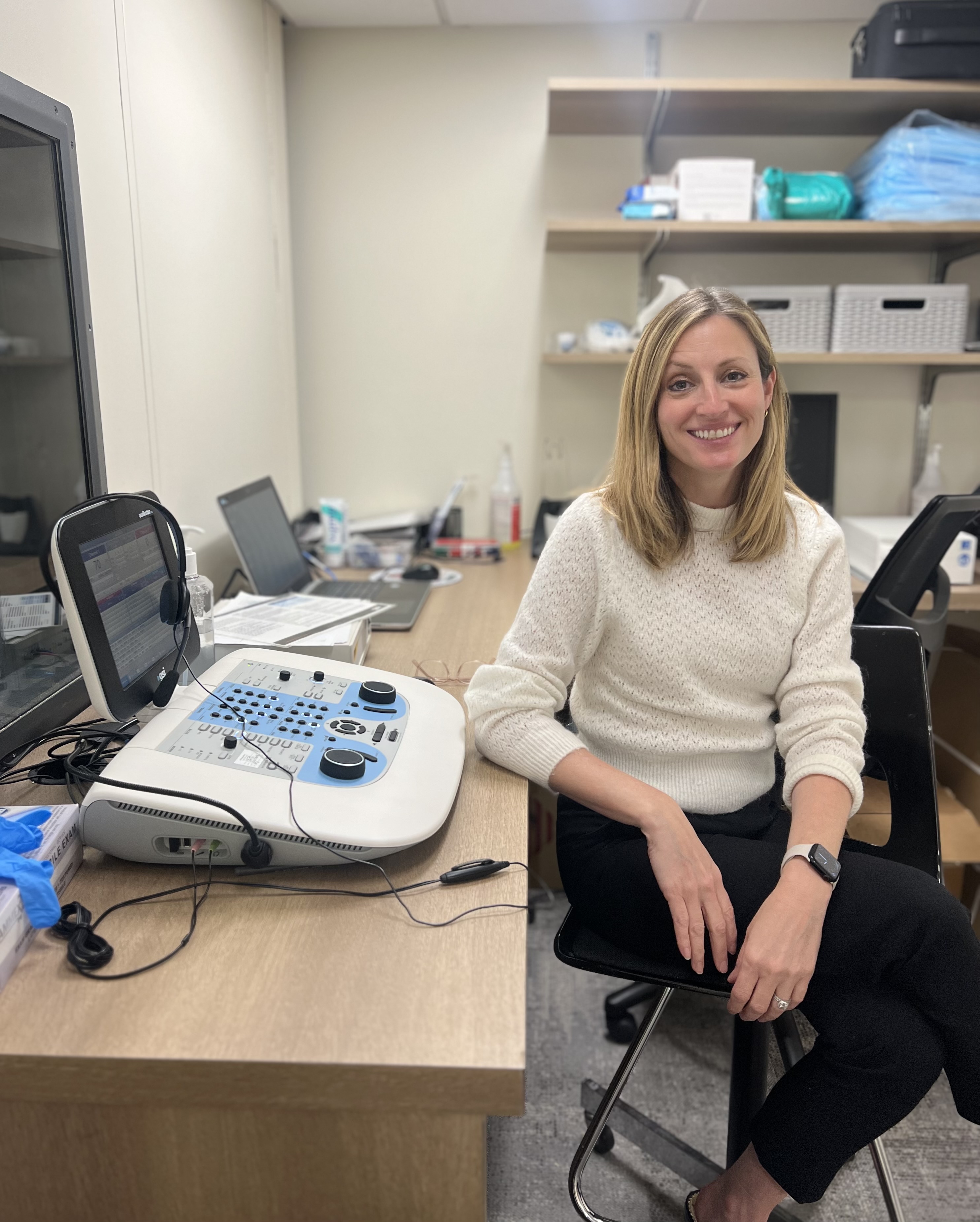
You will meet with Dr. Katie Brow, our research audiologist at the Neuroplasticity Research Center. You will be asked to complete questionnaires ranging from a case history to your own perception of your ability to hear in different listening situations. A vision and cognitive screening will also be conducted, followed by a comprehensive hearing evaluation in the hearing booth.
![]() During this evaluation, you will be asked to listen for tones and repeat words and/or sentences. You will then complete additional testing to evaluate your cognition. At the end, Dr. Brow will review your hearing results with you. She will let you know if you qualify for the study and your next steps in the process.
During this evaluation, you will be asked to listen for tones and repeat words and/or sentences. You will then complete additional testing to evaluate your cognition. At the end, Dr. Brow will review your hearing results with you. She will let you know if you qualify for the study and your next steps in the process.
Electroencephalograpy (EEG)
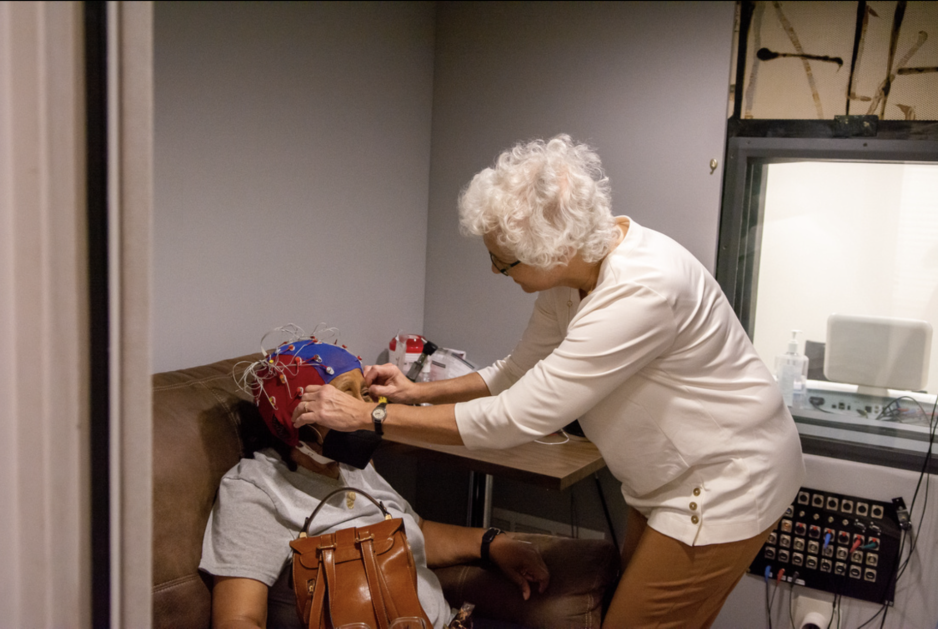
An electroencephalogram (EEG) is a test that measures electrical activity in the brain using small electrodes. The electrodes measure electrical signals (impulses) and the EEG machine records the impulses with lines (traces) that show wave patterns. For this study, you will be wearing a cortical cap that is gelled for each electrode. The gel used for the electrodes helps conduct impedance and get good data. For your EEG appointment, expect to get gel in your hair. It is a gooey material that easily washes out of your hair. Once you are wearing the cap and all the electrodes are secured, you will be asked to recline in a comfortable chair where you will be listening to different stimuli. While we record, it is important to stay relaxed and keep as still as possible to get the best results.
Magnetoencephalography (MEG)
MEG is a neuroimaging technique that our researchers use to measure brain activity.
How does it work? Researchers will place small sensors on your forehead that will be used to measure magnetic fields at the surface of the scalp. This does not involve any radiation and poses minimal safety risks. Throughout the MEG session, you will be in a quiet room, lying on your back with your head in a plastic helmet. During this time, you'll be listening to speech passages and answering questions while we monitor your brain. For a demonstration, please see our informational video below!
Magnetic Resonance Imaging (MRI)
MRI is another neuroimaging technique that our researchers use to produce an image of your brain.
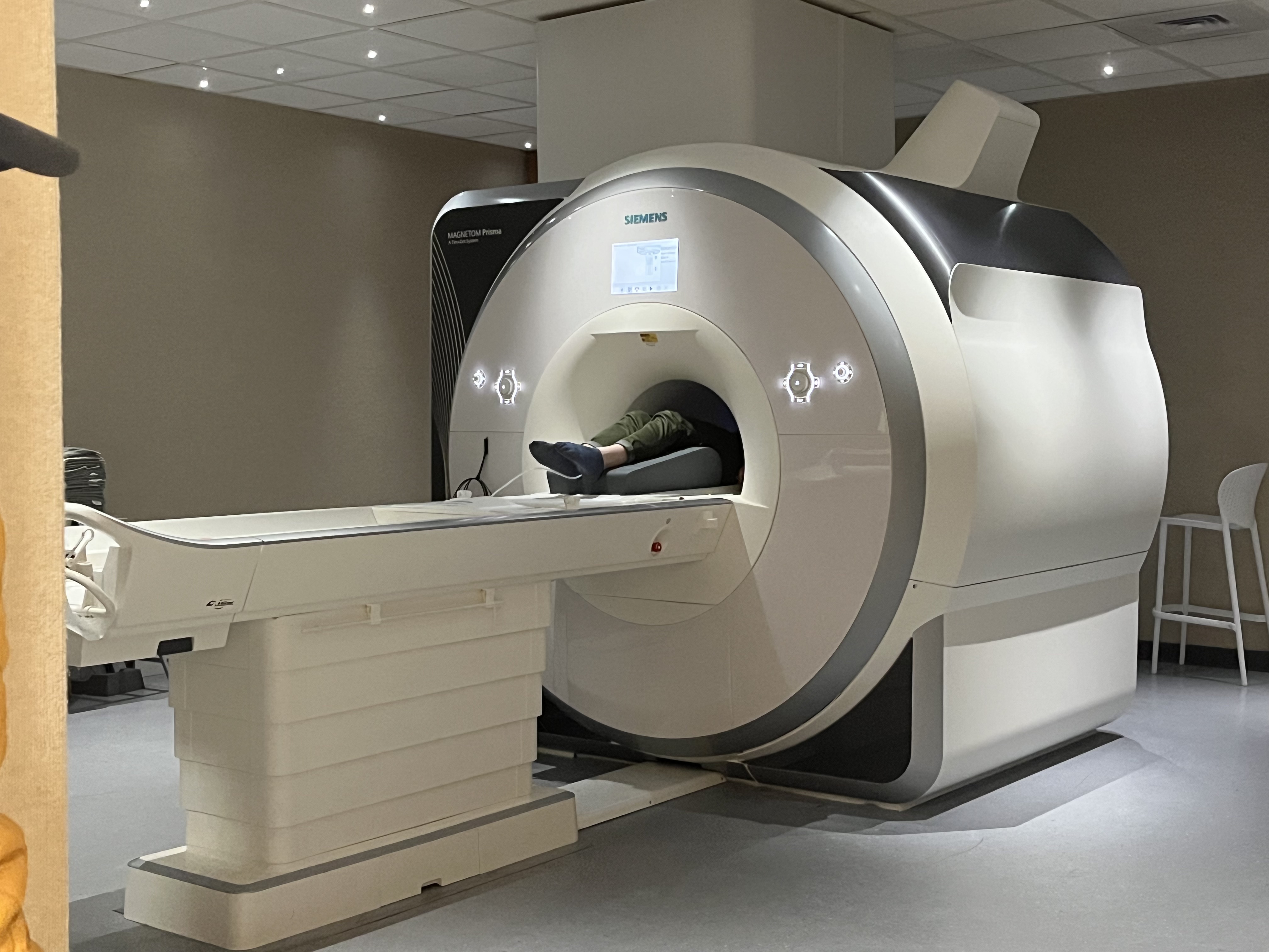
How does it work? MRI uses a strong magnetic field to produce images of an individuals brain or body. Similar to the MEG, it does not use radiation and poses minimal safety risks. Due to the strength of the magnetic field the MRI produces, it will be very important to not have any metal in your body or on your clothes. Before the session, you will go through a screening process to make sure all metal is removed. After, you will be asked to lay still on a flat table for about 30 minutes. You will not be performing any tasks during this session, so you can relax or watch a silent movie as we measure your brain. The two photos below show what the scanner will look like and what to expect during the session.
![]()


